
Scanning Electron Microscope (SEM) research, enlarged insect head, side view (3.4568)
Type: Photographic image
Description:Scanning Electron Microscope (SEM) research, enlarged insect, side view
Extent: 1 photograph
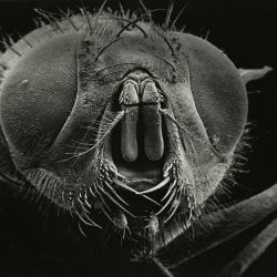
Scanning Electron Microscope (SEM) research, enlarged insect head, front view (3.4569)
Type: Photographic image
Description:Scanning Electron Microscope (SEM) research - enlarged insect head, front view
Extent: 1 photograph

Scanning Electron Microscope (SEM) research, enlarged insect head and partial body, side view (3.4570)
Type: Photographic image
Description:Scanning Electron Microscope (SEM) research, enlarged insect head and partial body, side view
Extent: 1 photograph
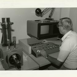
William Hess working at Scanning Electron Microscope (SEM) in laboratory (3.4571)
Date: 1988Type: Photographic image
Description:William Hess working at Scanning Electron Microscope (SEM) in laboratory
Extent: 1 photograph

Scanning Electron Microscope (SEM) research, enlarged insect body (3.4573)
Type: Photographic image
Description:Scanning Electron Microscope (SEM) research, enlarged insect body
Extent: 1 photograph

Scanning Electron Microscope (SEM) research, enlarged insect body (3.4574)
Type: Photographic image
Description:Scanning Electron Microscope (SEM) research, enlarged insect body
Extent: 1 photograph
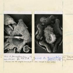
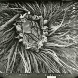
Scanning Electron Microscope (SEM) research, male flowers of (L) Acer pennsylvanicum and (R) Rhus aromatica (3.4576)
Creator: Crowley Jr., Webster R.
Type: Photographic image
Description:Scanning Electron Microscope (SEM) research, male flowers of (L) Acer pennsylvanicum and (R) Rhus aromatica
Lobed intrastaminal disk with remnants of stamens appear in between lobes
Flowers are not complete because parts were removed to show disk
Extent: 1 photograph

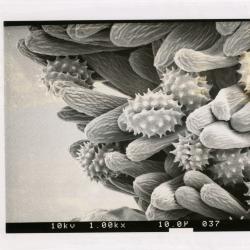
Scanning Electron Microscope (SEM) research, pollen grains of (L) Acer pseudoplatanus and (R) Rhus typhina (3.4578)
Creator: Crowley Jr., Webster R.
Type: Photographic image
Description:Scanning Electron Microscope (SEM) research, pollen grains of (L) Acer pseudoplatanus and (R) Rhus typhina
Shows the spaghetti-like ridges (striations) of the surface (exine) between the grooves (colpi)
Extent: 1 photograph
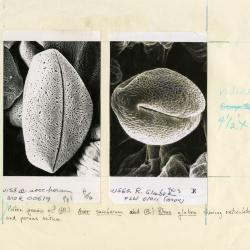
Scanning Electron Microscope (SEM) research, pollen grains of (L) Acer saccharum and (R) Rhus glabra (3.4580)
Creator: Crowley Jr., Webster R.
Type: Photographic image
Description:Scanning Electron Microscope (SEM) research, pollen grains of (L) Acer saccharum and (R) Rhus glabra
Shows porous nature of the veiny (reticulate) surface (exine) between the grooves (culpi)
Extent: 1 photograph

Scanning Electron Microscope (SEM) research, Croton (3.4582)
Date: March 1986Type: Photographic image
Description:Scanning Electron Microscope (SEM) research, Croton
Extent: 1 photograph
Scanning Electron Microscope (SEM) research, Populus deltoides, coma around seed (3.4583)
Date: March 1986Type: Photographic image
Description:Scanning Electron Microscope (SEM) research, Populus deltoides, coma around seed
Extent: 1 photograph

Scanning Electron Microscope (SEM) research, Gerbera (Compositae) African, Asian (3.4584)
Date: 1985Type: Photographic image
Description:Scanning Electron Microscope (SEM) research, Gerbera (Compositae) African, Asian
Extent: 1 photograph
Scanning Electron Microscope (SEM) research - SEM photo: Pollen grains of Tall Goldenrod (3.4585)
Date: 1986Creator: Hess, William J.
Type: Photographic image
Description:Scanning Electron Microscope (SEM) research - SEM photo: Pollen grains of Tall Goldenrod
Extent: 1 photograph

Scanning Electron Microscope (SEM) research - SEM photo: Pollen grain - flower of Tall Goldenrod (3.4586)
Date: 1986Creator: Hess, William J.
Type: Photographic image
Description:Scanning Electron Microscope (SEM) research - SEM photo: Pollen grain - flower of Tall Goldenrod
Extent: 1 photograph

Scanning Electron Microscope (SEM) research - SEM photo: Disk flower of Goldenrod (3.4587)
Date: 1986Creator: Hess, William J.
Type: Photographic image
Description:Scanning Electron Microscope (SEM) research - SEM photo: Disk flower of Goldenrod
Extent: 1 photograph

Scanning Electron Microscope (SEM) research - SEM photo: pollen (3.4588)
Type: Photographic image
Description:Scanning Electron Microscope (SEM) research - SEM photo: pollen
Extent: 1 photograph

Scanning Electron Microscope (SEM) research - SEM photo: insect (3.4589)
Type: Photographic image
Description:Scanning Electron Microscope (SEM) research - SEM photo: insect
Extent: 1 photograph

Scanning Electron Microscope (SEM) research - SEM photo: pollen from Rhus (3.4590)
Date: circa 1986Type: Photographic image
Description:Scanning Electron Microscope (SEM) research - SEM photo: pollen from Rhus
Extent: 1 photograph

Scanning Electron Microscope (SEM) research - SEM photo: Solidago altissima (3.4591)
Type: Photographic image
Description:Scanning Electron Microscope (SEM) research - SEM photo: Solidago altissima
Extent: 1 photograph

Scanning Electron Microscope (SEM) research - William Hess operating Scanning Electron Microscope (3.4595)
Type: Photographic image
Description:Scanning Electron Microscope (SEM) research - William Hess operating Scanning Electron Microscope
Extent: 1 photograph

Scanning Electron Microscope (SEM) research - William Hess holding stage of the SEM on which the disc flowers have been plated with gold (3.4596)
Date: 1986Creator: Kohout, John
Type: Photographic image
Description:Scanning Electron Microscope (SEM) research - William Hess holding stage of the SEM on which the disc flowers have been plated with gold
Extent: 1 photograph

Scanning Electron Microscope (SEM) research - William Hess placing the coated specimen on stage into the specimen chamber (3.4597)
Date: 1986Creator: Kohout, John
Type: Photographic image
Description:Scanning Electron Microscope (SEM) research - William Hess placing the coated specimen on stage into the specimen chamber
Extent: 1 photograph

Scanning Electron Microscope (SEM) research - William Hess covering the disc flower (on stage) ready for high voltage in a vacuum chamber (3.4598)
Date: 1986Creator: Kohout, John
Type: Photographic image
Description:Scanning Electron Microscope (SEM) research - William Hess covering the disc flower (on stage) ready for high voltage in a vacuum chamber
Extent: 1 photograph

Urban Vegetation Lab Research Staff group shot in laboratory - George Ware (seated), (standing L to R): Rick Hootman, Pat Kelsey, Jeff Mengler, Gary Watson, Mike Spravka (3.4599)
Date: circa 1987Creator: Irving, Gary
Type: Photographic image
Description:Urban Vegetation Lab Research Staff group shot in laboratory - George Ware (seated), (standing L to R): Rick Hootman, Pat Kelsey, Jeff Mengler, Gary Watson, Mike Spravka